operating room, petri dish

Medical & Biomedical Diagnostics

Raman spectroscopy is becoming more pervasive in biomedical diagnostics because of the demand for near real time and minimally invasive analysis at the point of care. Raman is an ideal technique for molecular fingerprinting and is sensitive to the chemical changes associated with disease.

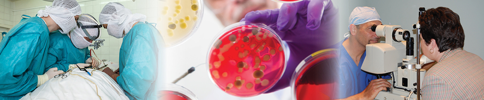
Biomedical science is one of the most exciting application areas of Raman spectroscopy because it can accomplish the diagnosis of disease at an early stage. Raman is a nondestructive technique that can be used for analysis including in vivo studies of tissues and cells, minimizing the need for tissue incisions. There is an increasing use of Raman for clinical applications because Raman is an ideal technique for molecular fingerprinting and is sensitive to the chemical changes associated with disease. Furthermore, components in the tissue matrix, principally those associated with water and bodily fluids, give a weak Raman response, thereby improving sensitivity to spectral signal changes associated with a diseased state. Raman analysis has been used for rapid in vivo screening for breast and cervical cancer, gastrointestinal tumors, cerebrospinal fluid related to spinal cord injuries, and many more medical diagnoses.

Biomedical Raman applications include: examination of biopsies, cytology, drug efficacy studies, histopathology, better defining surgical targets and the interface between malignant and healthy tissue, and treatment monitoring.

Some of the most active research areas are the analysis of abnormalities in tissue samples such as brain, arteries, breast, bone, cervix, embryonic media; and the identification of biomarkers for early stage detection of breast, cervical, and pancreatic cancer.

Raman has also been used to investigate blood disorders such as anemia and leukemia, as well as understanding cell growth in bacteria, phytoplankton, viruses and other micro-organisms.

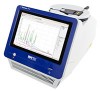
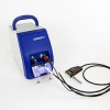
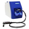
The portability of the i-Raman series instruments makes them ideal for rapid diagnostics of at the point of care. They can be used in vivo during surgery, or near the clinical settings for rapid screening of biopsy samples to identify malignant and benign tissue. The i-Raman series instruments are available with 532, 785, or 1064-nm excitation laser.

Products

i-Raman® Prime

High throughput, Highly Sensitive, High Resolution Raman System

i-Raman® EX

1064nm Fiber Optic Raman System

i-Raman® Plus

Highly Sensitive, High Resolution Fiber Optic Raman System

Testimonial

The B&W Tek Instrument [i-Raman Pro] was very easy to use. And in roughly one hour we were able to adapt to the unusual spatial requirements of performing Raman in a surgical suite. We were able to quantitatively 1) reproduce earlier results using 830 nm excitation in a rat model and 2) perform analogous experiment in pig model finding quantitative agreement with standard lab analysis of physically sampled Cerebrospinal Fluid.

— Prof. Joseph Chaiken, Professor of Chemistry, Syracuse University

Request a Quote or Information

Application Notes

The mysteries of our origins have intrigued humans since the beginning of time. In this application note, a Raman Spectrometer manufactured by B&W Tek was used in analysis to show feasibility of Raman for the ‘Exomars’ payload mission launched in 2018.
Read the full application note
Raman spectroscopy is a nondestructive and highly versatile technique for analysis of chemicals, both organic and inorganic. It is used in industry, bioscience, medical diagnosis, forensics, and many other areas.
Read the full application note
In this study, the feasibility of fluorescence background subtraction using a handheld spectrometer to differentiate several bacterial growth media, specifically in the presence and absence of bacteria was investigated. As SERS is often not practical for first responders and military personnel (because of time constraints, limited dexterity in personal protective equipment, and ease of use), a portable Raman spectrometer that utilizes a proprietary algorithm to subtract background fluorescence was tested. This baseline correction capability may allow for the interrogation of biologic materials that would otherwise “swamp” traditional handheld Raman instruments used in forensic analyses.
Read the full application note
A revolutionary method for identifying breast cancer in lymph nodes using Raman spectroscopy is being tested by researchers and surgeons at a UK hospital.
Read the full application note
This study shows that relatively inexpensive, portable Raman devices can be used to differentiate between malignant and healthy regions in breast tissue with a high degree of certainty. The boundary between malignant and benign breast tumors were evaluated using two commercial Raman systems: one operating with 1064 nm laser excitation source and the other 785 nm, wavelengths that are known to be capable of interrogating biological systems without target damage. The 1064 nm systems have a deeper penetration depth into tissue than 785 nm devices and often generate significantly less fluorescence. That is a significant advantage since fluorescence can easily mask the weaker Raman signal.In the initial evaluations, the authors employed the systems in microscopic mode and with acquisition setting s to make the total laser exposure equal for both systems. Multivariate statistical classification methods were applied to the spectral data using a minimal set of spectral bands identified and found to give reliable classification between healthy and malignant tissue using either the 1064 nm or 785 nm Raman systems.
Read the application note
Both Raman spectroscopy and surface enhanced Raman spectroscopy (SERS) are proving to be invaluable tools in the field of biomedical research and clinical diagnostics. The robust, compact, fit-for-purpose Raman spectrometer designs are appropriate for use in surgical procedures to help surgeons assess tumors and allow rapid decisions to be made. The two biomedical applications highlighted in this review demonstrate how this SERS technique can be an important part of the medical toolbox
Read the application note
Raman spectroscopy is becoming more pervasive in biomedical diagnostics because of the demand for near real time and minimally invasive analysis at the point of care. Raman is an ideal technique for molecular fingerprinting and is sensitive to the chemical changes associated with disease.
Read the application note
Tuberculosis (TB) remains one of the leading causes of death worldwide with 10.4 million new cases reported in 2015. In the same year, 1.8 million people died from TB even with the availability of a vaccine. The Bacillus Calmette-Guerin (BCG) vaccine is the only vaccine available against TB, despite its variable efficacy and its failure to control spread of the disease. Although the BCG vaccine provides protection against manifestations of TB in children, it is not able to provide reliable protection against adult pulmonary TB. Hence, there is an urgent need to develop a vaccine that can surpass the efficacy of the current BCG vaccine. In this study shows how Raman spectroscopy was used to develop a panel of biophysical methods that can monitor the adjuvantation process for a TB vaccine candidate and provide characterization information on formulated vaccine components without the need for desorption.
Read the application note
x