
Components of a Raman Spectrometer
In any Raman spectrometer, there are three main components – a sampling apparatus, an excitation source, and a detector. Although these components have come in varying forms over the years, advanced Raman instruments were developed using a laser as an excitation source, a spectrometer for the detector, and a fiber optic or a microscope for the sampling apparatus.
Raman spectroscopy is predicated on the ability to measure a shift in wavelength (or frequency) and, therefore, a monochromatic excitation source should be used. While a laser is usually the best excitation source, not all lasers can be used for Raman spectroscopy, so the laser frequency should be very stable and should not mode hop, as this will lead to errors in the Raman shift.
Also, a clean, narrow bandwidth laser should be used because the quality of the Raman peaks is directly influenced by the stability and sharpness of the excitation light source.
Wavelength is the final consideration when deciding which laser to use for a Raman spectrometer. From the previous section, it is clear that when the wavelength is shorter, the Raman signal becomes more powerful. However, as stated before, this is not the only consideration, particularly when it comes to dealing with organic molecules.
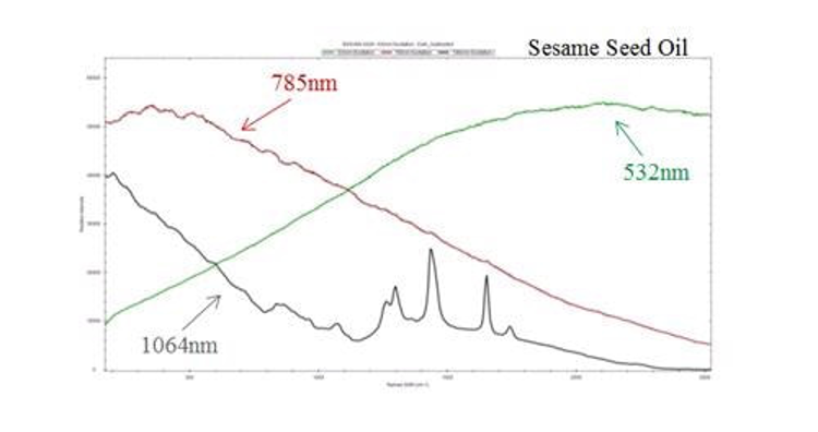
When excited by high energy (short wavelength) photons, most organic molecules will tend to fluoresce. Although fluorescence is usually considered to be a low light level process, it can still overwhelm the signal in the Raman spectrum (Figure 5). This is because the Raman effect contains a very small fraction (approximately 1 in 107) of the incident photons. Therefore, only visible lasers are used for inorganic materials such as carbon nanotubes.
In the case of organic molecules, laser wavelength should be shifted into the near infrared to reduce fluorescence without exceeding the CCD spectral detection limits. 785 nm diode lasers have become the industry standard because of their availability and the fact that they enable maximum fluorescence reduction without affecting the spectral range or resolution. A 532 nm laser is the best choice for increased sensitivity with inorganic molecules, as fluorescence is no longer an issue.
As discussed before, Raman scattering is extremely weak and needs long integration times to collect enough photons to measure a discernible signal. This makes it essential to use a TE cooled spectrometer to reduce the dark noise.
For weak Raman scatters or very low concentrations, a back-thinned CCD may be required to further increase the spectrometer‘s sensitivity. By etching the detector to just a few microns thick, the probability of an electron being reabsorbed as it travels through the detector based on Beer’s law is considerably reduced. This boosts the detector’s sensitivity from a maximum quantum efficiency of 35% to more than 90%.
Due to the highly selective nature of Raman spectra, they may include closely spaced peaks that may need to be resolved depending on the application. This can be achieved using a high resolution spectrometer. Usually, standard spectrometer configurations are meant for 785 nm and 532 nm laser excitation wavelengths, but custom excitation wavelengths are also available.
These spectrometers can provide a range of configurations that are exclusively designed for high resolution and wide spectral range. Standard spectral ranges are available from as low as 65 cm-1 (filter dependent) to as high as 4000 cm-1, with a spectral resolution as fine as 3 cm-1.
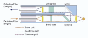 Whenever a sample is measured, the only effective way of directing the laser light to the sample, collecting the Raman scatter, and directing it to a spectrometer is to use a fiber optic probe. A Raman probe must be able to direct and focus the monochromatic excitation source (usually a laser) to the sample, collecting the scattered light, and then directing it to the spectrometer. A typical design for a Raman probe is shown in Figure 6.
Given that a pure signal is very important to Raman spectroscopy, a narrow band-pass filter is placed in the optical path of the excitation source before it reaches the sample. As the Raman effect is very weak, the signal should be collected at a 0° angle normal to the sample. This results in interference from Rayleigh scattering, and therefore the collected signal should be filtered through a long pass filter before it is directed to the spectrometer.
The flexibility of fiber optics allows the probe to be taken to a solid sample and also enables it to be immersed in slurries or liquids in both process and lab environments (for kinetic measurements in real time). Fiber optics can also be coupled to cuvette holders, microscopes, and a host of sampling accessories.